Bones In Leg Diagram : Guide To Knee Joint Anatomy - The talus the weight of your body is transferred from the tiba to the talus.
byAdmin-
0
Bones In Leg Diagram : Guide To Knee Joint Anatomy - The talus the weight of your body is transferred from the tiba to the talus.. The lower limb contains 30 bones. Check spelling or type a new query. The tibia, commonly known as the 'shin bone', is the largest and most medial of the two.you can palpate its anterior border when you run your finger down the anterior aspect of your leg. 12 photos of the bones leg diagram picture. This diagram depicts diagram leg bones anatomy.human anatomy diagrams show internal organs, cells, systems, conditions, symptoms and sickness information and/or tips for healthy living.
The bones of the leg are the femur, tibia, fibula and patella.the foot bones shown in this diagram are the talus, navicular, cuneiform, cuboid, metatarsals and calcaneus. The lower leg extends from the knee to the ankle. Maybe you would like to learn more about one of these? Inflammation of navicular bone and/or bursa. The bones together make up the hip.
Ankle Anatomy Muscles And Ligaments from embed.widencdn.net The proximal portion of the tibia is tibial plateau which acts as a cusp for the knee, the distal portion tapers into the medial malleoli and the concave surface which articulates with the talus at the ankle joint. 12 photos of the bones leg diagram picture. Bones of right thigh and leg. The talocrual joint is made up of three main bones. The distal ends of the radius and ulna bones articulate with the hand bones at the junction of the wrist, which is formally known as the carpus. Bones in leg diagram color the leg on the left side. The pubis, ischium, and ilium together constitute the pelvis while the thigh bone is the femur. The lower leg is comprised of two bones, the tibia and the smaller fibula.
Schema de legs bones diagram diagram showing bones inside human leg ready to jump stock file skeleton of a cat diagram ver 2 svg disposition of rotator cuff muscles diagram.
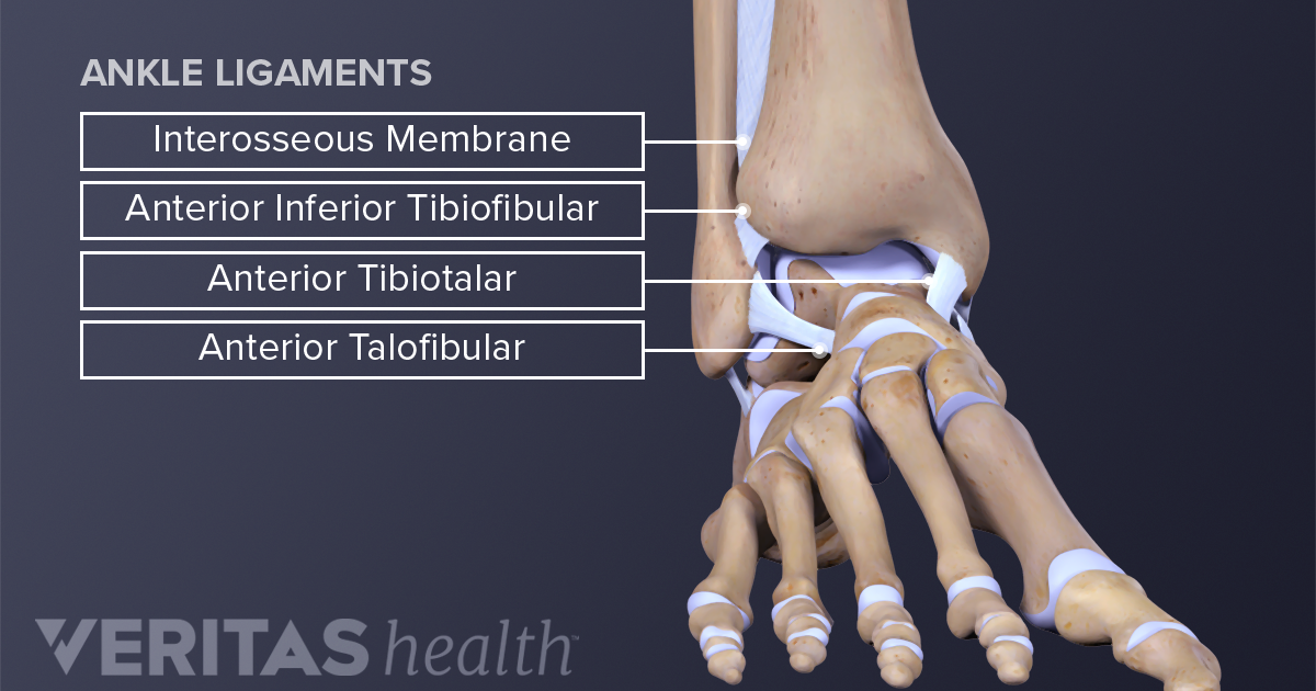
Tibia and fibula the tibia and fibula are two long bones that run parallel to each other, forming the scaffold of the leg and providing attachment points for many muscles. The bones of the foot are divided into three groups. The forearm contains two major bones. The ligament joining the two bones of the lower leg (tibia and fibula), called the syndesmotic ligament, is injured. These are the femur, patella, tibia, fibula, tarsal bones, metatarsal bones, and phalanges (see figure 6.51). The femur is the single bone of the thigh. The stifle joint connects the femur, which is the dog thigh bone, to the tibia and fibula, the lower leg bones, and the patella,the canine equivalent to the knee cap. Your legs are two of your most important body parts. Bone diagram forehead (frontal bone) nose bones (nasals) cheek bone (zygoma) upper jaw (maxilla) lower jaw (mandible) breast bone (sternum) upper arm bone (humerus) lower arm bone (ulna) thigh bone (femur) collar bone (clavicle) toe bones (phalanges) ankle bones (tarsals) kneecap (patella) shin bone Four quadriceps muscles are present in front of the knee which help in straightening the leg from the knee. This diagram depicts diagram leg bones anatomy.human anatomy diagrams show internal organs, cells, systems, conditions, symptoms and sickness information and/or tips for healthy living. Pin on medical websites we like. The knee joint is the largest joint in the body and is primarily a hinge joint, although some sliding and rotation occur.
We did not find results for: The lower leg extends from the knee to the ankle. The lower limb contains 30 bones. We did not find results for: The technical term for a dog knee is the stifle joint.
Leg Picture Image On Medicinenet Com from images.medicinenet.com Related posts of diagram of leg bones inside of arm muscle and bone. Ankle & lower leg anatomy. Inside of arm muscle and bone 12 photos of the inside of arm muscle and bone , bone The tibia and fibula are the bones of the lower leg. Pin on medical websites we like. Numbered one through five the bone that sits behind the big toe is no. The bones of the leg are the femur, tibia, fibula and patella.the foot bones shown in this diagram are the talus, navicular, cuneiform, cuboid, metatarsals and calcaneus. Bones of right thigh and leg.
The tibia and fibula form the ankle joint with the talus,.
It is likely that abnormal biomechanical stresses are the basis for the disease. Learn about leg bone anatomy, including general leg anatomy, the names of major leg bones, and the location and function of each bone. Bone diagram forehead (frontal bone) nose bones (nasals) cheek bone (zygoma) upper jaw (maxilla) lower jaw (mandible) breast bone (sternum) upper arm bone (humerus) lower arm bone (ulna) thigh bone (femur) collar bone (clavicle) toe bones (phalanges) ankle bones (tarsals) kneecap (patella) shin bone Inflammation of navicular bone and/or bursa. Another bone that is part of the lower leg and the knee joint is called the fibula.this is a bone located on the lateral, or outer part, of the lower leg and is more commonly known as the calf bone. Check spelling or type a new query. The smaller lateral bone of the lower leg. The lower leg is comprised of two bones the tibia and the smaller fibula. The thigh bone, or femur, is the large upper leg bone that connects the lower leg bones (knee joint) to the pelvic bone (hip joint). Schema de legs bones diagram diagram showing bones inside human leg ready to jump stock file skeleton of a cat diagram ver 2 svg disposition of rotator cuff muscles diagram. These bones are firmly attached with the help of muscles and tendons which also save the bones from injury. Numbered one through five the bone that sits behind the big toe is no. The tibia and fibula are the bones of the lower leg.
Learn about leg bone anatomy, including general leg anatomy, the names of major leg bones, and the location and function of each bone. We did not find results for: Leg bone anatomy diagram diagram of human leg human anatomy human leg bones anatomy stock photo download image now anatomy of the knee central coast orthopedic medical group Check spelling or type a new query. These bones are firmly attached with the help of muscles and tendons which also save the bones from injury.
Lower Extremity Anatomy Bones Muscles Nerves Vessels Kenhub from thumbor.kenhub.com The knee joint is the largest joint in the body and is primarily a hinge joint, although some sliding and rotation occur. The lower leg is comprised of two bones, the tibia and the smaller fibula. This diagram depicts diagram leg bones anatomy.human anatomy diagrams show internal organs, cells, systems, conditions, symptoms and sickness information and/or tips for healthy living. Its lower end helps create the knee joint. The tibia is much larger than the fibula and bears almost all of the body's weight. The medial, larger bone of the lower leg. The pubis, ischium, and ilium together constitute the pelvis while the thigh bone is the femur. The proximal portion of the tibia is tibial plateau which acts as a cusp for the knee, the distal portion tapers into the medial malleoli and the concave surface which articulates with the talus at the ankle joint.
The stifle joint connects the femur, which is the dog thigh bone, to the tibia and fibula, the lower leg bones, and the patella,the canine equivalent to the knee cap.
The hip itself is a ball and socket joint, much like the shoulder.the structures necessary to create this joint are the socket, the joint capsule, muscle, ligaments, and the neck. The bones together make up the hip. The femur is the single bone of the thigh. Degenerative disease, similar to arthritis. When you stand or walk, all the weight of your upper body rests on them. The distal ends of the radius and ulna bones articulate with the hand bones at the junction of the wrist, which is formally known as the carpus. 12 photos of the bones leg diagram picture. A high ankle sprain causes pain and swelling similar to a. The forearm contains two major bones. These bones are firmly attached with the help of muscles and tendons which also save the bones from injury. Inflammation of navicular bone and/or bursa. The talocrual joint is made up of three main bones. We did not find results for: